
Optics for Intelligent Measurement (OPTIM) develops optical sensing and imaging technologies for healthcare, biotechnology and other challenging environments. We combine optical fibres, meta-optics and miniaturised devices with computational imaging and AI. Our aim is to sense and image where conventional technologies cannot, connecting sensor design and fabrication with calibration, reconstruction, interpretation and validation.
Click a theme or topic in the map to explore our research and papers.
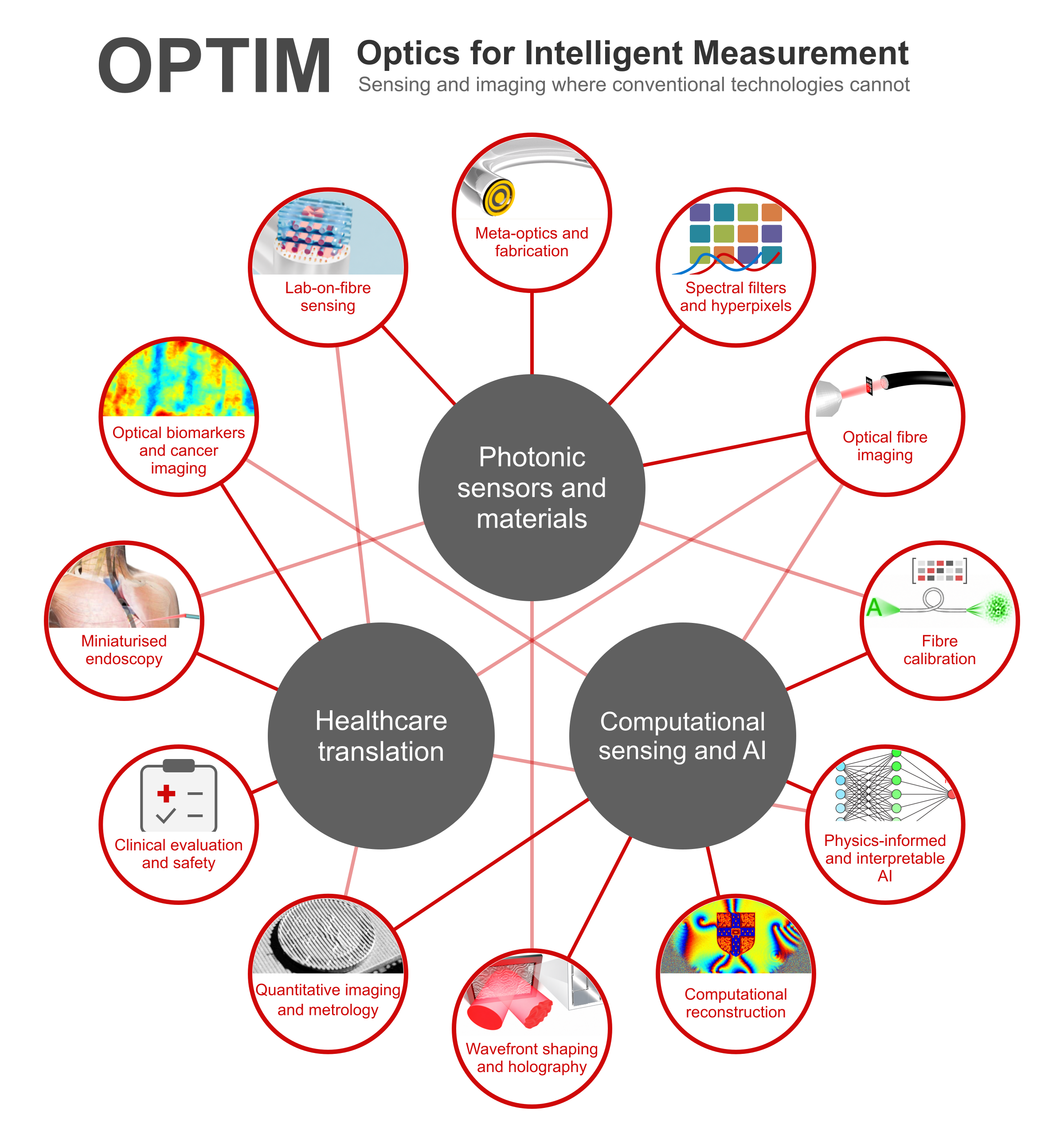
From optical devices to useful measurements
Explore the map to see how our research connects.
Photonic sensors and materials
Designing and fabricating the devices that collect and shape light.
Computational sensing and AI
Calibrating instruments and recovering reliable, interpretable information.
Healthcare translation
Developing and evaluating technologies for minimally invasive imaging and sensing.
Photonic sensors and materials
We develop optical fibres, meta-optics and miniaturised devices that control how light interacts with a sample. Our work combines scalable fabrication of planar lenses on fibre tips with application-specific spectral filter arrays. These technologies connect sensor design and manufacture to computational reconstruction and compact biomedical imaging.
Explore this theme
Computational sensing and AI
We develop calibration, reconstruction and machine-learning methods that turn complex measurements into useful information. Examples include neural-network recovery of fibre transmission matrices, high-speed wavefront shaping and interpretable analysis of phonon microscopy data. Physical models and experimental validation help us understand what a method learns and where it can be used.
Explore this theme
Healthcare translation
We work towards minimally invasive imaging and sensing for earlier disease detection. Our research spans miniaturised quantitative endoscopy, label-free tissue contrast and clinical studies of endoscopy procedures. We evaluate technologies in phantoms and biological samples, and work with clinical collaborators to address the practical requirements of their use in healthcare.
Explore this theme
Meta-optics and fabrication
We fabricate thin optical structures that shape light at the tips of hair-thin fibres. Our scalable transfer process for single- and multi-layer planar lenses supports both tightly focused and extended-depth beams, with preliminary demonstrations of lateral imaging and optical coherence tomography. This approach connects planar manufacturing to miniature imaging probes. Related work explores holographic metasurfaces whose response changes with the surrounding medium.
Connected themes
Spectral filters and hyperpixels
We design spectral filters around the information an imaging task needs. Hyperpixels combine multiple optical resonators within each pixel to tailor its spectral response, improving discrimination between target spectra in our experiments. They build on our single-step fabrication of bespoke multispectral filter arrays. These compact devices offer a route to application-specific spectral imaging in miniature instruments.
Connected themes
Optical fibre imaging
We use optical fibres to carry images through narrow spaces, supporting the development of hair-thin endoscopes. By measuring and correcting how a fibre transforms light, we recover amplitude, phase and polarisation images through a fibre bundle. Our work also demonstrates controlled intensity and phase patterns through a multimode fibre, including image formation away from the fibre tip. Probe hardware, calibration and reconstruction are designed together.
Connected themes
Fibre calibration
Fibre bending and temperature changes can scramble an image, making calibration essential. We developed a reflection-based framework using metasurface reflector stacks and neural networks that recover transmission matrices from simulated single-ended measurements, cross-validated with experimentally measured matrices. Related studies address interferometer phase drift and measurement noise. Together, these methods work towards reliable imaging when the far end of the fibre is inaccessible.
Connected themes
Physics-informed and interpretable AI
We combine machine learning with knowledge of the measurement process. Our fibre-calibration networks use a loss function that accounts for an ambiguity in optical phase. In phonon microscopy, multi-task conditional networks reduce differences between experimental batches while classifying cells and recovering signals that can be interpreted physically. Published conference work extends physics-informed AI to fibre calibration from partial measurements.
Connected themes
Computational reconstruction
We develop algorithms that recover meaningful images from distorted or indirect measurements. This includes reconstruction of optical vector fields for endoscopic imaging and neural-network mapping of tissue absorption and scattering from structured-light images. In the latter work, synthetic data provide known ground truth and realistic geometries; experiments combining synthetic and measured training data test how the method transfers to physical samples.
Connected themes
Wavefront shaping and holography
We control the spatial structure of light for imaging, projection and propagation through complex media. Our work includes fast, light-efficient wavefront shaping with a MEMS phase-only modulator and phase-retrieval methods for generating structured fields through multimode fibres. We also contribute practical computational tools, including HoloGen for high-speed hologram generation.
Connected themes
Quantitative imaging and metrology
We model how illumination, sample shape and light scattering affect quantitative measurements. Our open-source 3D simulation framework for spatial frequency domain imaging tests realistic geometries, including the inside of tubular organs, and helps identify calibration requirements before a system is built. The same modelling approach supplies synthetic training data for optical-property reconstruction, linking instrument design to computational analysis.
Connected themes
Clinical evaluation and safety
We work with clinicians to evaluate practical questions around endoscopic procedures. Our studies have measured aerosol and droplet generation during gastrointestinal endoscopy, investigated air filtration in endoscopy rooms and compared aerosol production during Cytosponge and conventional upper gastrointestinal endoscopy. This work complements optical technology development with experience of measurement in real clinical settings.
Connected themes
Miniaturised endoscopy
We develop small endoscopes that capture more information than conventional white-light images. Our dual-wavelength spatial frequency domain imaging prototype measures absorption and scattering in tissue-mimicking phantoms using miniature fibre illumination and a micro-camera. Complementary work on hyperspectral endoscopy demonstrated spectral imaging in phantoms and excised tissues, including imaging during freehand motion. These studies establish capabilities needed for future clinical evaluation.
Connected themes
Optical biomarkers and cancer imaging
We investigate tissue properties that could reveal disease without added labels. Phase and polarisation imaging through optical fibres revealed early tumours in excised mouse oesophageal tissue that were not apparent in amplitude-only images. Hyperspectral imaging distinguished spectral profiles in normal and pathological excised human tissue. Related phonon microscopy research connects cell classification to interpretable mechanical features, including adhesion.
Connected themes
Lab-on-fibre sensing
We integrate optical functionality onto fibre tips to create compact probes for imaging and sensing. Transferred planar optics provide a fabrication route for miniature devices with tailored beam shapes. Published conference work also explores an optical-fibre refractive-index sensor modified with a gold nanodisc metasurface. This direction links nanostructured materials with measurements close to the sample.